So you’re pregnant and you keep hearing about all these different types of ultrasounds you’re gonna get. Dating scan, anatomy scan, transvaginal, 3D – it’s like there’s a whole menu of options and nobody bothered to explain what any of them actually are or why you need them.
I remember sitting in my first OB appointment and my doctor started talking about scheduling various scans throughout my pregnancy. I’m nodding along like I know what she’s talking about, but inside I’m thinking “wait, how many ultrasounds am I actually getting here?” Turns out, way more than I expected, and each one serves a totally different purpose.
Let me walk you through every type of ultrasound you’re likely to encounter during these nine months. No medical school required, just straight talk about what each scan does and when you’ll be getting it.
The two basic ultrasound methods
Before we get into the specific types of scans, you gotta understand the two main ways they actually do ultrasounds. Everything else builds from these two basic approaches.
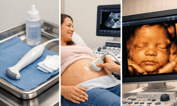
Transabdominal ultrasound
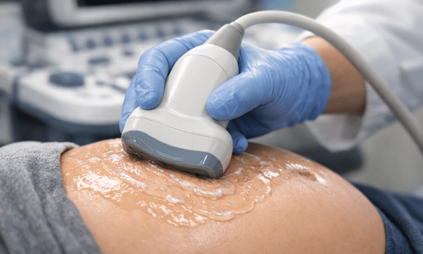
This is what most people picture when they think “ultrasound.” You lie back on the exam table, the tech squirts that cold gel all over your belly, and then moves a device called a transducer across your skin. The gel helps the sound waves travel from the transducer through your skin and into your uterus.
The transducer sends out high-frequency sound waves that bounce off your baby and internal structures, then converts those echoes into images on a screen. It’s completely safe – no radiation involved, which is why doctors can use it as often as needed throughout pregnancy.
You’ll need a moderately full bladder for most transabdominal ultrasounds, especially early in pregnancy. The full bladder pushes your uterus up and out of your pelvis, making it easier to see. Trust me, finding that sweet spot between “full enough” and “about to pee my pants” is an art form.
Transvaginal ultrasound
Now this one surprises some first-time moms. A transvaginal ultrasound uses a slender wand-shaped transducer that’s inserted into your vagina. I know, sounds weird if you’ve never had one, but it’s really not as uncomfortable as it sounds.
Why would they do this instead of just using the regular belly scan? Because in early pregnancy, your uterus is still tucked down in your pelvis, and a transvaginal probe can get way closer to the action. The images are clearer and more detailed, especially before about 10 or 11 weeks.
The probe is covered with a disposable protective sheath and lubricated gel. You can actually insert it yourself if that makes you more comfortable, or the sonographer will do it gently. The whole thing takes maybe 10 or 15 minutes, and for this one you’ll want an empty bladder.
Early pregnancy scans
Viability or dating scan (6-10 weeks)
This is usually your first official peek at your baby. The main goals here are confirming that you’re actually pregnant, making sure the pregnancy is in the right place (in your uterus, not ectopic), seeing that heartbeat for the first time, and getting an accurate due date.
Most of the time this is done transvaginally because your baby is tiny – we’re talking the size of a blueberry or raspberry at this point. The abdominal approach just can’t get close enough to see clearly yet.
What you’ll see is pretty basic. A small gestational sac, a yolk sac that’s feeding your baby before the placenta takes over, and if you’re far enough along, a flickering heartbeat that’ll probably make you cry. I definitely did.
The sonographer will measure the crown-rump length to date your pregnancy. This measurement is crazy accurate this early on, usually within 3 to 5 days of your actual conception date.
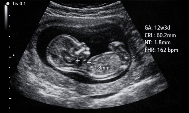
First trimester screening (11-14 weeks)
This scan, sometimes called the nuchal translucency (NT) scan, happens toward the end of your first trimester. It’s checking for chromosomal abnormalities like Down syndrome by measuring the fluid at the back of your baby’s neck.
The NT measurement combined with blood work gives you a risk assessment, not a diagnosis. If the results show higher risk, your doctor might recommend more definitive testing like NIPT (non-invasive prenatal testing) or amniocentesis.
At this scan, your baby actually looks like a baby for the first time instead of a blob. You might see them moving around, waving their little arms. It’s wild. They’ll also confirm your due date one more time and count fingers and toes if your baby cooperates with their positioning.
Second trimester scans
Anatomy scan (18-22 weeks)
This is the big one. The anatomy scan, also called the level 2 ultrasound or the 20-week scan, is the most detailed ultrasound of your pregnancy. Plan on being there for at least 30 to 45 minutes, sometimes longer if your baby’s being stubborn and won’t move into good positions.
The sonographer is checking everything – and I mean everything. Brain, spine, heart chambers and vessels, kidneys, bladder, stomach, all four limbs, the placenta, amniotic fluid levels, and your cervix. They’re taking tons of measurements and screenshots to make sure all your baby’s organs are forming correctly.
This is also when most people find out the sex if they want to know. The tech will usually ask if you want to find out before they get to that part of the exam.
Here’s something nobody tells you – sometimes they can’t see everything in one session. Maybe the baby’s curled up in a weird position or the placenta is blocking the view of something. Don’t freak out if they ask you to come back in a week or two for a follow-up. It’s super common and usually everything turns out fine once they get a better angle.
Growth scans (as needed)
Some women get additional ultrasounds in the second or third trimester to check on baby’s growth. This isn’t routine for everyone – it’s usually recommended if you have certain risk factors like gestational diabetes, high blood pressure, or if your baby was measuring small or large at the anatomy scan.
These scans measure your baby’s head, abdomen, and femur to estimate their weight and make sure they’re growing appropriately. They’ll also check your amniotic fluid levels and take a look at blood flow through the umbilical cord using something called Doppler ultrasound.
Third trimester scans
Biophysical profile (BPP)
If you make it to or past your due date, or if your doctor has any concerns about your baby’s wellbeing late in pregnancy, you might get a biophysical profile. This combines an ultrasound with a non-stress test.
The ultrasound portion checks four things – fetal breathing movements, fetal body movements, fetal tone (like flexing and extending), and amniotic fluid volume. Each component gets a score of either 0 or 2, and they add them all up. A score of 8 or 10 is reassuring.
The non-stress test monitors your baby’s heart rate and any contractions you might be having. They’re looking for the heart rate to accelerate when the baby moves, which is a sign that everything’s good.
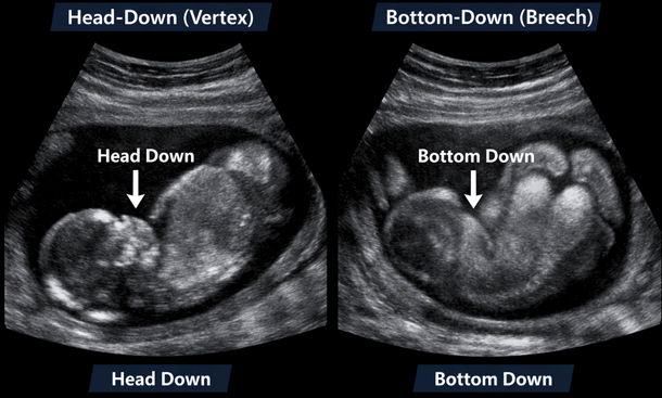
Presentation check
Toward the end of pregnancy, around 36 weeks or so, your doctor might do a quick ultrasound just to confirm which way your baby’s positioned. Are they head down and ready to go, or are they breech with their butt or feet pointing toward your cervix?
This scan takes like two minutes. They’re not doing detailed measurements or anything, just confirming position so they can plan for delivery appropriately.
Special ultrasound types
3D and 4D ultrasounds
Okay so regular ultrasounds give you 2D images – flat, black and white pictures. 3D ultrasounds use the same sound wave technology but the computer processes the data differently to create three-dimensional images of your baby’s face and body.
4D is basically 3D in real time, so you see a 3D video instead of still images. You can watch your baby yawn, suck their thumb, make faces – it’s pretty amazing.
Here’s the thing though – these aren’t typically part of routine prenatal care. They’re mostly done at specialty boutique ultrasound places, and you pay out of pocket. Some people love them and think they’re worth every penny. Others think they’re a waste of money when you’re gonna meet your baby face-to-face soon enough anyway.
If you do want one, the best time is usually between 26 and 32 weeks when your baby has enough fat to fill out their features but isn’t so big that they’re all squished up with no room to move.
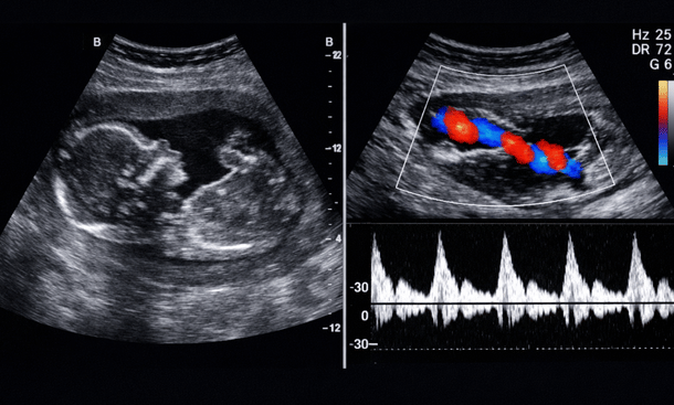
Doppler ultrasound
This isn’t really a separate type of scan – it’s a technique that can be added to a regular ultrasound. Doppler measures blood flow through vessels, particularly the umbilical cord, the vessels in your baby’s brain, and sometimes the blood flow to your uterus.
Your doctor might order this if there are concerns about your baby’s growth or if you have certain medical conditions. The Doppler creates color images showing which direction blood is flowing and how fast, which helps assess whether your baby’s getting adequate oxygen and nutrients.
You’ll hear a whooshing sound during a Doppler – that’s the sound of blood flowing through the vessels. It’s different from the heartbeat sound you hear with a regular fetal Doppler at your prenatal visits.
What about all those elective ultrasounds
You’ve probably seen ads for places that do “keepsake” ultrasounds where you can bring your whole family, get a bunch of pictures and videos, and make it this whole event. These places aren’t medical facilities and the people doing the scans aren’t necessarily trained sonographers.
The FDA actually recommends against getting ultrasounds purely for entertainment or keepsake purposes. Not because ultrasounds are dangerous – they’re not – but because untrained operators might miss something important, or worse, tell you something’s wrong when it’s not and scare you unnecessarily.
If you want extra ultrasounds beyond what your doctor orders, talk to your OB about it. Some practices offer additional scans in-office for reassurance, especially if you’ve had a previous loss or are just really anxious.
How to prepare for different scan types
The prep for your ultrasound depends on which type you’re having. For early transabdominal scans, you’ll need that full bladder I mentioned – usually they tell you to drink about 32 ounces of water an hour before and don’t pee.
For transvaginal ultrasounds, empty your bladder right before. Wear comfortable clothes that are easy to pull up or take off. You’ll probably be given a sheet to drape over your lower half.
For the anatomy scan and later ultrasounds, bladder fullness doesn’t matter as much. Just show up comfortable and ready to lie still for a while. Bring your partner or support person if you want – most places allow one person in the room with you.
When you might need more ultrasounds than average
Most low-risk pregnancies involve two or three ultrasounds total – one in the first trimester, the anatomy scan, and maybe one more toward the end. But certain situations mean you’ll be seeing that ultrasound machine a lot more often.
Multiples pregnancies get way more monitoring. High-risk conditions like gestational diabetes, preeclampsia, or placenta previa require regular growth and position checks. Previous pregnancy complications often mean closer surveillance this time around.
IVF pregnancies typically get more early scans. If you’ve had previous losses, your doctor might offer more frequent ultrasounds in the first trimester for reassurance.
The bottom line on ultrasound types
Look, all these different scans can feel overwhelming when you’re trying to keep track of what’s happening when. But each one serves a specific purpose in monitoring your baby’s development and making sure everything’s progressing normally.
You don’t need to memorize what happens at each scan. Your doctor’s office will tell you what to expect and how to prepare for each appointment. What matters is understanding that these aren’t just opportunities to see cute pictures of your baby – though that’s definitely a nice bonus – they’re important medical assessments.
The technology has come so far that we can catch and address issues early that would’ve gone undetected a generation ago. Try to see each ultrasound as a chance to check in on your baby’s development and make sure they’re growing strong and healthy. If you want to dive deeper into interpreting what you see at each of these scans, our comprehensive guide to understanding pregnancy ultrasound results covers everything from measurements to normal findings at every stage.
And if you’re ever confused about the numbers and abbreviations you see on your ultrasound reports during any of these scans, you’ll want to read our breakdown of how to read ultrasound pictures so you can actually understand what you’re looking at on that screen instead of just squinting and nodding.

As an author at Felyro.com, I create actionable content on pregnancy tracking, offering practical tools, tips, and insights that empower mothers-to-be to stay informed and confident throughout their pregnancy.