
You just got back from your ultrasound and you’re sitting there with this report full of numbers and medical terms, trying to figure out if everything’s actually okay or if you should be worried. Your doctor said “everything looks normal” but what does that even mean? Normal compared to what?
I’ve been in that exact spot, staring at measurements and percentiles, Googling every single number like some kind of obsessed detective. With my first pregnancy, I convinced myself something was wrong because one measurement was in the 35th percentile and another was in the 65th. Turns out that’s completely fine – babies aren’t required to be exactly average in every single category.
So let me break down what “normal” actually means when it comes to ultrasound results, what kind of variations are totally expected, and what might actually warrant a follow-up conversation with your doctor.
What normal actually means on an ultrasound
Here’s the thing about normal – it’s not one specific number. It’s a range, and that range is actually pretty wide for most measurements. When doctors say your results are normal, they mean your baby’s measurements fall within expected parameters for their gestational age.
Those parameters come from studying thousands and thousands of pregnancies and creating growth charts that show what’s typical. But typical doesn’t mean identical. Just like adults come in different sizes and proportions, so do babies in utero.
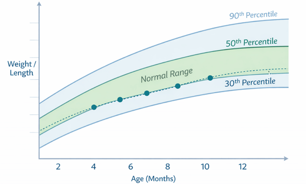
Most measurements are considered normal if they fall between the 10th and 90th percentile. That means 80 percent of babies at that gestational age fall within this range. If your baby’s consistently measuring around the 25th percentile for everything, that’s normal. If they’re consistently around the 75th percentile, that’s also normal. They’re just on different growth curves.
What doctors look for is consistency and appropriate growth over time. A baby measuring in the 40th percentile at one scan and then the 38th percentile at the next scan is fine – that’s basically the same. A baby who drops from the 60th to the 20th percentile between scans might need closer monitoring.
Normal first trimester findings
In the first trimester, normal means seeing a gestational sac in the right place – inside your uterus, not in a fallopian tube or anywhere else. The sac should be round or oval, with clear borders.
By about 5 and a half to 6 weeks, you should see a yolk sac inside the gestational sac. This looks like a small ring and it’s feeding your baby before the placenta takes over. Seeing the yolk sac is a good sign that the pregnancy is developing.
The fetal pole – that’s your actual baby at this stage – should be visible by around 6 weeks. It looks like a small white curve or blob attached to the yolk sac. And here’s the big one – the heartbeat. By 6 to 7 weeks, you should see that little flicker of the heart beating.
Normal heart rate in early pregnancy is between 110 and 170 beats per minute, though it’s usually on the higher end, like 150 to 170. If the heart rate is in this range and you can see a clear, strong flicker, that’s exactly what you want.
Crown-rump length (CRL) measurements in the first trimester should correlate with your gestational age. At 8 weeks, normal CRL is around 1.6 cm. By 12 weeks, it’s usually around 5.4 cm. There’s some variation, but if the measurement matches up with your dates within a few days, you’re good.
The nuchal translucency (NT) measurement, done between 11 and 14 weeks, should be less than 3 mm for most pregnancies. This measurement combined with blood work helps assess risk for certain chromosomal conditions. If it’s under 3 mm, that’s considered a reassuring finding.
What’s normal at the anatomy scan
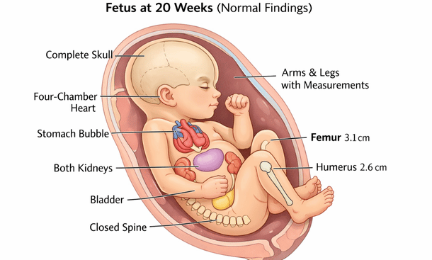
The anatomy scan around 20 weeks is when they really get into the details. This is where a lot of women start freaking out because there’s so much being measured and evaluated.
For the head, a normal biparietal diameter (BPD) at 20 weeks is around 4.7 cm, give or take. Head circumference (HC) should be around 17.5 cm. These measurements increase as pregnancy progresses, and there’s a range of normal for each week.
Abdominal circumference (AC) at 20 weeks is typically around 15 to 16 cm. This one can vary quite a bit based on whether your baby just swallowed some fluid or if they’re all curled up.
Femur length (FL) at 20 weeks should be approximately 3.4 cm. Again, there’s normal variation here. Some babies have longer legs, some have shorter ones, just like adults.
The four-chamber view of the heart should show two atria and two ventricles that are roughly equal in size. The heart should be positioned on the left side of the chest and should be beating rhythmically. Normal fetal heart rate in the second and third trimesters is between 120 and 160 beats per minute.
Both kidneys should be visible and roughly the same size. You should be able to see the stomach as a small dark bubble in the abdomen – this shows your baby’s swallowing amniotic fluid. The bladder should also be visible as a dark circle in the pelvis.
The spine should show all the vertebrae lined up with no gaps or protrusions. The skull should be completely closed with no openings. All four limbs should be present with normal bone development.
Normal amniotic fluid levels
The amount of fluid surrounding your baby is measured in different ways depending on when in pregnancy you are. Too much or too little can both be concerns, but there’s a pretty wide range of normal.
One way they measure is the amniotic fluid index (AFI), which adds up the deepest pockets of fluid in four quadrants of your uterus. Normal AFI is between 5 and 25 cm. Less than 5 is oligohydramnios (too little), more than 25 is polyhydramnios (too much).
Another method is the single deepest pocket (SDP) or maximum vertical pocket (MVP). Normal is considered to be between 2 and 8 cm.
Your fluid levels change throughout pregnancy. They increase until around 34 to 36 weeks and then start to decrease slightly toward your due date. This is totally normal.
If your fluid levels are just slightly outside the normal range, your doctor might just want to recheck in a week or two. Hydration can affect fluid levels, so sometimes drinking more water actually helps if levels are borderline low.
Normal placenta position and appearance
The placenta can be positioned in different spots and still be totally normal. Fundal (at the top), anterior (front), posterior (back), or on either side – all of these are normal locations.
What you don’t want is a placenta that’s covering your cervix, called placenta previa. Even if the placenta is low early in pregnancy, it usually moves up as your uterus grows. If it’s still covering the cervix after 28 weeks, that’s when it becomes more of a concern.
Normal placenta thickness is roughly the same as the gestational age in millimeters until about 36 weeks. So at 20 weeks, around 20 mm thick is typical. After 36 weeks, it stays around 35 to 40 mm thick.
The texture should look relatively uniform and smooth. The grade or maturity of the placenta increases as pregnancy progresses, and that’s normal. You might see calcifications or areas that look a bit different, and often that’s just normal aging of the placenta.
Normal variations that look weird but aren’t problems
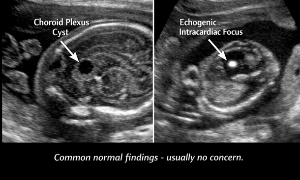
There are some findings that sound scary when you first hear them but are actually pretty common and usually not concerning.
Choroid plexus cysts are small fluid-filled spaces in the brain that show up on some second trimester ultrasounds. They’re present in about 1 to 2 percent of pregnancies and almost always go away on their own by the third trimester. When they occur alone without other abnormalities, they’re not associated with problems.
Echogenic intracardiac focus (EIF) is a bright spot in the heart that shows up in about 5 percent of pregnancies. It’s usually just a normal variation in how calcium is deposited in the heart muscle. It doesn’t affect heart function and typically doesn’t mean anything is wrong.
Echogenic bowel means the baby’s intestines appear brighter than usual on ultrasound. This can be totally normal, or it can sometimes be associated with other conditions. If it’s the only finding and not severe, it’s often just a normal variant.
A two-vessel umbilical cord (missing one artery) occurs in less than 1 percent of pregnancies. Many babies with this are completely healthy, though your doctor might do some additional monitoring.
Dilated renal pelvis (slight swelling in the kidney) is pretty common and often resolves on its own. Mild cases usually don’t cause any problems.
When measurements don’t all match up
Sometimes your baby measures in the 70th percentile for head size but the 30th percentile for femur length. Before you panic, let me tell you this is super common and usually not a problem.
Babies, just like adults, have different body proportions. Maybe you have a tall family and your partner’s family is shorter. Your baby might have a longer torso and shorter legs – that’s genetics, not a problem.
What doctors look for is whether the measurements are roughly proportional. If the head is measuring way bigger than everything else, they might want to take a closer look to make sure there’s no fluid buildup or other issues. If the abdomen is measuring much smaller than the head and limbs, they might check placental function to make sure baby’s getting enough nutrition.
Small variations between percentiles are totally normal. We’re talking about differences of 20 or 30 percentile points being usually fine. It’s when you see major discrepancies – like head in the 90th percentile and abdomen in the 10th percentile – that additional evaluation might be needed.
Normal fetal position throughout pregnancy
Your baby’s position changes constantly throughout pregnancy, and there’s a wide range of normal.
In the first and second trimesters, babies flip around all the time. They have plenty of room to move, so position doesn’t really matter yet. You might see them head down at one scan and breech at the next – completely normal.
By the third trimester, especially after 34 weeks or so, most babies settle into a head-down position. About 96 percent of babies are head-down by the time you go into labor. That’s the ideal position for birth, but plenty of babies don’t get there until the very end.
Breech position (bottom or feet first) is common earlier in the third trimester. If your baby’s still breech at 36 or 37 weeks, your doctor might discuss options like external cephalic version (trying to turn the baby) or planning for a C-section.
Transverse position (sideways) is less common but also usually resolves on its own before labor. Babies have a way of finding their way into the right position when it’s time.
Growth patterns and what’s considered appropriate
Appropriate growth means your baby is getting bigger over time at a steady rate. They should be following their own growth curve, even if that curve is at the lower or higher end of normal.
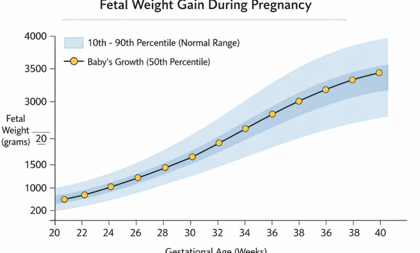
In the second trimester, babies grow about 1.5 cm per week in length. Weight gain accelerates in the third trimester, with babies typically gaining about half a pound per week in the last month or so.
If your baby was measuring in the 40th percentile at 20 weeks and is still in the 35th to 45th percentile at 28 weeks, that’s good consistent growth. If they dropped from the 60th to the 20th percentile, your doctor would want to investigate why the growth slowed down.
Estimated fetal weight (EFW) becomes more important in the third trimester. Normal weight at 28 weeks is around 2.2 pounds, at 32 weeks around 3.7 pounds, at 36 weeks around 5.8 pounds, and at 40 weeks around 7.5 pounds. But remember, these are estimates with a margin of error of about a pound in either direction.
Small for gestational age (SGA) means measuring below the 10th percentile. Large for gestational age (LGA) means above the 90th percentile. Both can be totally normal – some babies are just small or large – but they might warrant extra monitoring to make sure everything’s okay.
Normal findings you might not expect
There are some things that are completely normal but might surprise you if nobody mentioned them beforehand.
It’s normal to see your baby’s gender organs on ultrasound. If you’re having a boy, you might see the penis and scrotum pretty clearly. If you’re having a girl, you might see the labia. This is just part of the anatomy scan and helps confirm gender if you want to know.
Hiccups show up on ultrasound as rhythmic little jumps or bounces. They’re totally normal and show that your baby’s diaphragm is developing and practicing breathing movements.
You might see your baby making breathing movements even though they’re not actually breathing air. This is practice for after birth and it’s a good sign of healthy development.
Babies swallow amniotic fluid constantly – that’s why you can see the stomach on ultrasound. They also pee in the amniotic fluid. This is normal and healthy. The fluid is constantly being recycled.
Your baby might look like they’re sucking their thumb or fingers on the ultrasound. Lots of babies do this in utero. It’s adorable and completely normal.
Understanding Doppler results
If your doctor orders Doppler ultrasound to check blood flow, here’s what normal looks like.
Umbilical artery Doppler should show continuous forward flow throughout the cardiac cycle. The resistance should be low, meaning blood flows easily from the placenta to your baby. As pregnancy progresses, the resistance normally decreases even more.
Middle cerebral artery (MCA) Doppler checks blood flow to your baby’s brain. Normal is low resistance with good forward flow. If the resistance is too high or flow patterns look abnormal, it might indicate your baby isn’t getting enough oxygen.
The ductus venosus is a blood vessel in your baby’s liver area. Normal flow should be forward throughout the heartbeat cycle. Abnormal flow patterns here can indicate heart problems or other issues.
Your doctor might also check uterine artery Doppler to assess blood flow to your placenta. Normal is low resistance with good flow. High resistance can be associated with preeclampsia or growth restriction risks.
If all the Doppler studies look normal, that’s very reassuring that your baby’s getting good blood flow and oxygen.
What to do if something’s borderline
Sometimes you’ll get results that are technically within normal range but on the edge. Maybe your fluid is at 5.5 when normal is 5 to 25, or your baby’s measuring in the 12th percentile when normal starts at 10.
First, don’t panic. Borderline doesn’t mean abnormal. It just means your doctor wants to keep a closer eye on things.
Ask your doctor what the plan is. Usually it’s just repeat ultrasounds in a week or two to make sure things aren’t getting worse. Sometimes they might recommend increasing your water intake, resting more, or making other small changes.
Follow the monitoring schedule your doctor recommends. If they want to recheck in a week, don’t push it to two weeks because you’re busy. These follow-ups are important for catching any changes early.
Keep track of your baby’s movements. If you notice a decrease in movement, call your doctor right away, even if you just had a normal ultrasound.
Ask questions if you don’t understand something. Your doctor should be able to explain what they’re watching for and why they’re not more concerned about borderline findings.
The bottom line on normal results
Normal ultrasound results mean your baby is growing and developing the way they should be for their gestational age. It doesn’t mean every measurement is exactly at the 50th percentile or that everything looks identical to other babies at the same stage.
Your baby is unique, and their measurements will reflect that. As long as growth is consistent, measurements are roughly proportional, and your doctor isn’t expressing concerns, you can feel confident things are on track.
Try not to obsess over every single number on the report. I know it’s hard – trust me, I’ve been there with my calculator and Google at 2 AM trying to figure out if my baby’s femur being 0.3 mm shorter than average meant something. It didn’t.
Focus on the overall picture. Is your baby growing? Is the heart beating normally? Are all the organs developing? Is there good amniotic fluid? Those are the things that matter.
And remember, ultrasounds are screening tools, not diagnostic tools. If something looks unusual, your doctor will order additional testing to get more information. Most of the time, those follow-up tests come back completely normal. For a comprehensive look at understanding all aspects of your pregnancy ultrasound results – from reading the images to interpreting measurements to knowing what types of scans you’ll get – our complete guide has everything you need to navigate your ultrasound appointments with confidence
If you’re still confused about what all those abbreviations and numbers on your ultrasound report actually mean, or if you want to understand exactly what’s being measured and why, take a look at our detailed questions to ask your doctor about ultrasound results so you can make the most of your next appointment and get all your concerns addressed.

As an author at Felyro.com, I create actionable content on pregnancy tracking, offering practical tools, tips, and insights that empower mothers-to-be to stay informed and confident throughout their pregnancy.